Chapter 17: Sound Waves - Lesson 2: Properties of Sound and Their Perception
The Human Ear
Hold down the T key for 3 seconds to activate the audio accessibility mode, at which point you can click the K key to pause and resume audio. Useful for the Check Your Understanding and See Answers.
Understanding how humans hear is a complex subject involving the fields of physiology, psychology and acoustics. In this part of Lesson 2, we will focus on the acoustics (the branch of physics pertaining to sound) of hearing. We will attempt to understand how the human ear serves as an astounding transducer, converting sound energy to mechanical energy to a nerve impulse that is transmitted to the brain. The ear's ability to do this allows us to perceive the pitch of sounds by detection of the wave's frequencies, the loudness of sound by detection of the wave's amplitude and the timbre of the sound by the detection of the various frequencies that make up a complex sound wave.
The ear consists of three basic parts - the outer ear, the middle ear, and the inner ear. Each part of the ear serves a specific purpose in the task of detecting and interpreting sound. The outer ear serves to collect and channel sound to the middle ear. The middle ear serves to transform the energy of a sound wave into the internal vibrations of the bone structure of the middle ear and ultimately transform these vibrations into a compressional wave in the inner ear. The inner ear serves to transform the energy of a compressional wave within the inner ear fluid into nerve impulses that can be transmitted to the brain. The three parts of the ear are shown below.
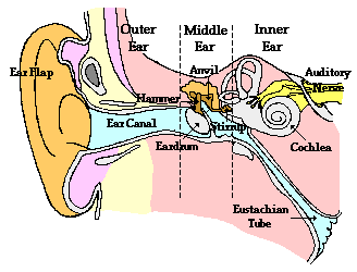
The Outer Ear
The outer ear consists of an earflap and an approximately 2-cm long ear canal. The earflap provides protection for the middle ear in order to prevent damage to the eardrum. The outer ear also channels sound waves that reach the ear through the ear canal to the eardrum of the middle ear. Because of the length of the ear canal, it is capable of amplifying sounds with frequencies of approximately 3000 Hz. As sound travels through the outer ear, the sound is still in the form of a pressure wave, with an alternating pattern of high and low pressure regions. It is not until the sound reaches the eardrum at the interface of the outer and the middle ear that the energy of the mechanical wave becomes converted into vibrations of the inner bone structure of the ear.
The Middle Ear
The middle ear is an air-filled cavity that consists of an eardrum and three tiny, interconnected bones - the hammer, anvil, and stirrup. The eardrum is a very durable and tightly stretched membrane that vibrates as the incoming pressure waves reach it. As shown below, a compression forces the eardrum inward and a rarefaction forces the eardrum outward, thus vibrating the eardrum at the same frequency of the sound wave.
Being connected to the hammer, the movements of the eardrum will set the hammer, anvil, and stirrup into motion at the same frequency of the sound wave. The stirrup is connected to the inner ear; and thus the vibrations of the stirrup are transmitted to the fluid of the inner ear and create a compression wave within the fluid. The three tiny bones of the middle ear act as levers to amplify the vibrations of the sound wave. Due to a mechanical advantage, the displacements of the stirrup are greater than that of the hammer. Furthermore, since the pressure wave striking the large area of the eardrum is concentrated into the smaller area of the stirrup, the force of the vibrating stirrup is nearly 15 times larger than that of the eardrum. This feature enhances our ability of hear the faintest of sounds. The middle ear is an air-filled cavity that is connected by the Eustachian tube to the mouth. This connection allows for the equalization of pressure within the air-filled cavities of the ear. When this tube becomes clogged during a cold, the ear cavity is unable to equalize its pressure; this will often lead to earaches and other pains.
The Inner Ear
The inner ear consists of a cochlea, the semicircular canals, and the auditory nerve. The cochlea and the semicircular canals are filled with a water-like fluid. The fluid and nerve cells of the semicircular canals provide no role in the task of hearing; they merely serve as accelerometers for detecting accelerated movements and assisting in the task of maintaining balance. The cochlea is a snail-shaped organ that would stretch to approximately 3 cm. In addition to being filled with fluid, the inner surface of the cochlea is lined with over 20 000 hair-like nerve cells that perform one of the most critical roles in our ability to hear. These nerve cells differ in length by minuscule amounts; they also have different degrees of resiliency to the fluid that passes over them. As a compressional wave moves from the interface between the hammer of the middle ear and the oval window of the inner ear through the cochlea, the small hair-like nerve cells will be set in motion. Each hair cell has a natural sensitivity to a particular frequency of vibration. When the frequency of the compressional wave matches the natural frequency of the nerve cell, that nerve cell will resonate with a larger amplitude of vibration. This increased vibrational amplitude induces the cell to release an electrical impulse that passes along the auditory nerve towards the brain. In a process that is not clearly understood, the brain is capable of interpreting the qualities of the sound upon reception of these electric nerve impulses.